


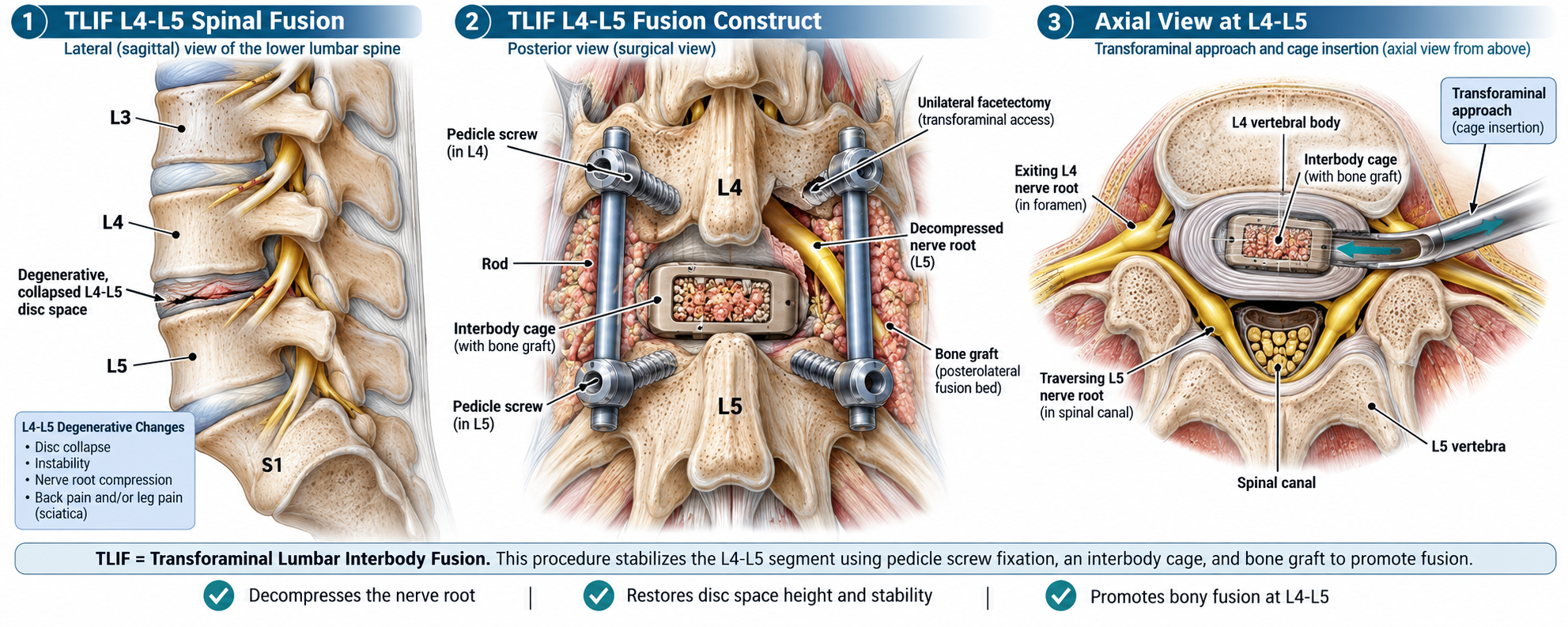
I wanted to share this graphic because it gives a clear look at what happens during a TLIF at L4-L5.
TLIF stands for Transforaminal Lumbar Interbody Fusion. The procedure is designed to stabilize the segment between the L4 and L5 vertebrae while also creating more room for compressed nerves.
The first view shows the lower lumbar spine from the side and highlights a degenerative, collapsed L4-L5 disc space. Changes at this level can contribute to instability, nerve compression, back pain, and pain that travels into the leg.
The second view shows the fusion construct from the back. Pedicle screws are placed into L4 and L5 and connected with rods to stabilize the segment. An interbody cage filled with bone graft is positioned between the vertebrae, while additional bone graft can be placed around the back of the spine to support fusion.
The third view looks down at L4-L5 from above and shows the transforaminal approach. This pathway allows the surgeon to reach the disc space from one side, decompress the affected nerve structures, remove damaged disc material, and insert the interbody cage.
The goal is not simply to place hardware. The screws and rods provide immediate mechanical stability while the bone graft gradually develops into a solid fusion between L4 and L5. The procedure can also restore disc-space height and reduce pressure on the affected nerve root.
This illustration does a good job of showing the three major parts of the operation: decompression of the nerve, stabilization with screws and rods, and the use of an interbody cage and bone graft to promote fusion.
As with any spinal procedure, the exact surgical approach and recovery can vary from one person to another, so this is best viewed as a general educational overview rather than individual medical advice.